Embryology - Biology 104, Spring 2006 - Albert Harris and Corey Johnson
NOTES FOR FOURTH LECTURE: Jan 18, 2006, by Albert Harris
Differentiation and Symmetry
What does anatomy consist of?
Differentiated cell types ~ 250 in humansArranged in certain spatial patterns
Specific examples include red blood cells, liver parenchyma cells, endothelial cells (that line blood vessels) and many others.
Cell differentiation is caused by selective expression of genes:
and this is mostly controlled at the level of transcription
(making messenger RNA or not, for that gene)
Some genes are "turned on" = messenger RNA is transcribed
-
remainder of genes are turned off; = not transcribed
only made in one or a few cell types
versus "Housekeeping" genes housekeeping proteins
; made in all or nearly all differentiated cell types
Differentiation according to position in embryo
Also active cell movements to new positions
Morphogenetic cell movements,
including gastrulation, neurulation and other events.
Also VERY important: Programmed cell death "Apoptosis"
Cells can self-destruct by activation of cytoplasmic enzymes that digest proteins.
This is what happens to the tails of tadpoles and the cells in the spaces between your fingers,
and there are many other uses of apoptosis (including maybe new cures for cancer, heart attacks and strokes! This will be explained later in the course.)
Cells reach all these differentiated states by means of certain branching pathways that are evolutionarily rather conservative, and very nearly the same for all vertebrates! (invertebrates have somewhat different versions of this branching pathway; for example they do have gastrulation but don't have neurulation, nor somites, notochord etc.)
Start with fertilized egg: One Cell
1) Series of rapid divisions ("cleavage stages")
produces blastula stage
(the equivalent stage is called the "blastocyst" in mammals, where it is somewhat special)
2) These cells rearrange and later begin to differentiate
More than half of the cells that start out on the surface move to interior
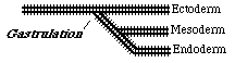
These inward movements are first set of cell rearrangements & are called "Gastrulation"
The cells left on the surface in gastrulation are called the ectoderm
Those that moved to the interior are called endoderm and mesoderm
These are called "the 3 primary germ layers "
(NOT in the sense of disease-causing germs)
ectoderm -> nervous system & the outer layer of skin
mesoderm -> muscles, bones, heart, blood vessels, kidneys, etc.
& the inner layer of the skin
endoderm -> lining of digestive tract, liver, lungs, some other organs
![]()
![]()
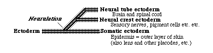
Neurulation is the subdivision of the ectoderm, separating the part that becomes brain & spinal cord away from the part that becomes skin. This is usually done by folding into a tube, and fusing the edges together (but not in teloest fish!)
![]()
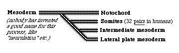
The mesoderm also undergoes morphogenetic rearrangements that subdivide it into...
-
* notochord
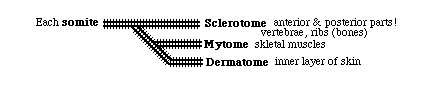
* somites
-> sclerotome -> skeleton* intermediate mesoderm
-> dermatome -> inner layer of skin
-> myotome -> voluntary muscles
-> pronephros, & pronephric duct (male sex ducts)* lateral plate mesoderm
-> mesonephros (kidney during development)
-> metanephos (kidney after birth)
-> visceral layer
with the coelomic cavity between these layers -> somatic layer
heart
female sex ducts
Other branching sequences could be drawn to illustrate the origin of the organs and cell types of the endoderm. But you see the pattern. Some kind of genetic switches control the progressive levels of specialization.
The remainder of this lecture dealt with Symmetry, a the key concept for understanding shape formation
Please continue with the following NOTES ON SYMMETRY.