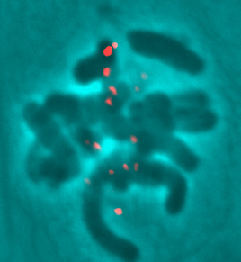
Howell, B.J., Hoffman, D.B., Fang, G., Murray, A.W., and Salmon, E.D. 2000. Visualization of Mad2 dynamics at kinetochroes, along spindle fibers, and at spindle poles in living cells. Journal of Cell Biology. 150(6): 1233-1250.

Time-lapse of Fluorescent Mad2 in Living, Mitotic PtK1 Cell:
Fluorescence and Phase Contrast Montage
Color Overlay of Fluorescence and Phase Contrast

Laser Photobleaching of Fluorescent Mad2 Protein on an Unattached Kinetochore in a Prometaphase PtK1 Cell

Visualization of the Release of a Mad2 Particle from the Kinetochore and Transport of the Particle to the Spindle Pole

Visualization of Mad2 Depletion at the Kinetochores and the Subsequent Loss of Mad2 at the Proximal Spindle Pole

Coming Soon... Mad2 "Streaks"


Canman, J.C., Hoffman, D.B., and Salmon, E.D. 2000. The role of pre- and post-anaphase microtubules in the cytokinesis phase, or C-phase, of the cell cycle. Current Biology. in press.

Control untreated cell injected with anti-Mad2 antibodies
          by Julie C. Canman
                Notice that the cell enters anaphase before properly alligning the chromosomes

Nocodazole-treated cell injected with anti-Mad2 antibodies- Example 1
Nocodazole-treated cell injected with anti-Mad2 antibodies- Example 2
Nocodazole-treated cell injected with anti-Mad2 antibodies- Example 3
          by Julie C. Canman
                Notice entry into anaphase is visible by the physical separation of the sister chromatids. Around the time of anaphase onset, the cortex becomes contractile about 10 min after antibody injection, and then ceases contraction about one hour after antibody injection. This indicates a distinct period of the cell cycle during which the cortex is contractile followed by the cessation of this contraction. This period is called C-phase for the cytokinetic phase of the cell cycle.

Nocodazole-treated cell injected with anti-Mad2 antibodies to induce anaphase, then released from nocodazole in C-phase...No cytokinesis
          by Julie C. Canman
                Washout is seen by the absence of images and lasts for ~5 min.

Nocodazole-treated cell injected with anti-Mad2 antibodies to induce anaphase, then released from nocodazole in C-phase...CYTOKINESIS
          by Julie C. Canman
                Washout is seen by the absence of images and lasts for ~5 min. Notice the cell underwent cytokinesis upon nocodazole washout!


Salmon Lab Home|Research Focus|Current Members|Salmon Lab Movies|Publications|Protocols|Members of Olde|Related Web Links


The Salmon Lab
University of North Carolina at Chapel Hill
Department of Biology
607 Fordham Hall,  CB#3280
Chapel Hill, NC  27599

To contact us:
Phone: (919) 962-2354
Fax: (919) 962-1625
Email: jccanman@email.unc.edu