Embryology - Biology 104, Spring 2006 - Albert Harris and Corey Johnson
LECTURE - March 27, 2006, by Albert Harris
More organs that form from Ectoderm
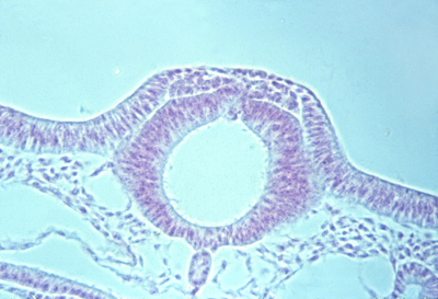
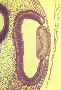
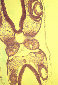
In this section of a chicken embryo, you can actually see the neural crest separating from the neural tube below and the somatic ectoderm above and to each side. (Also notice how small the notochord is, under the neural tube).
Somatic Ectoderm -->
-
epidermis (the outer layer of the skin
hair
feathers
scales in reptiles, birds & mammals
ameloblasts (form enamel of teeth)
{Except shark scales are very much like teeth! I don't know if they are formed by a combination of neural crest & epidermis?}
Somatic Ectoderm--> Stomodeum (infolding that forms the mouth)
Also formed from cells of the stomodeum are ameloblasts
These secrete the outer, enamel, layer of teeth
They are induced to form by the odontoblasts, which develop from neural crest cells.
This is an example of reciprocal induction, since both cell types apparently induce development of the other.
(neither can develop without the other)
Prof. William Koch of the UNC med school did some of the most important early research on this reciprocal induction)
*******************************************************************************************
hair: formed entirely by somatic ectodermal cells (epidermis)
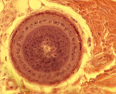
But each hair's formation is induced by a tightly bunched-together mass of mesodermal mesenchymal cells, called a dermal papilla. These control where hair form, and (apparently) the diameters of each hair.
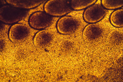
Feathers in birds are also induced to form by mesodermal papillae. Also scales in reptiles, birds and mammals, also each have a dermal (mesoderm) placode.
The locations of feathers and scales is EXTREMELY regular, almost crystalline in pattern. It is a long standing unsolved problem to discover the mechanisms that controls where they form
Hair locations are usually much less regular, except for certain hairs like the vibrissae above the upper lips of dogs.
Also, the next time you are petting a dog, notice that the formation of regular-sized hairs seems to be inhibited for a short distance around each of the vibrissae.