C. elegans Gastrulation:
an overview
"Internalization of multiple cells during C. elegans gastrulation
depends on common cytoskeletal mechanisms
but different cell polarity and cell fate regulators"
Jessica R. Harrell and Bob
Goldstein
|
| C. elegans gastrulation presents an opportunity to examine (1) mechanisms by which cells internalize from the surfaces of embryos, and (2) the extent to which diverse mechanisms may be used by distinct cells undergoing morphogenesis — by dozens of cells that are internalized at distinct times within a single organism. We identified 66 cells that are internalized in C. elegans gastrulation, many of which were not known previously to gastrulate. We found that diverse cell fate and cell polarity regulators control common mechanisms of morphogenesis in C. elegans. The results highlighted the variety of developmental patterning mechanisms that can be associated with common cytoskeletal mechanisms in the morphogenesis of an animal embryo. |
|
||||||||||||||||||||||||||||||||||||||||||||||||||||||||||||||||||
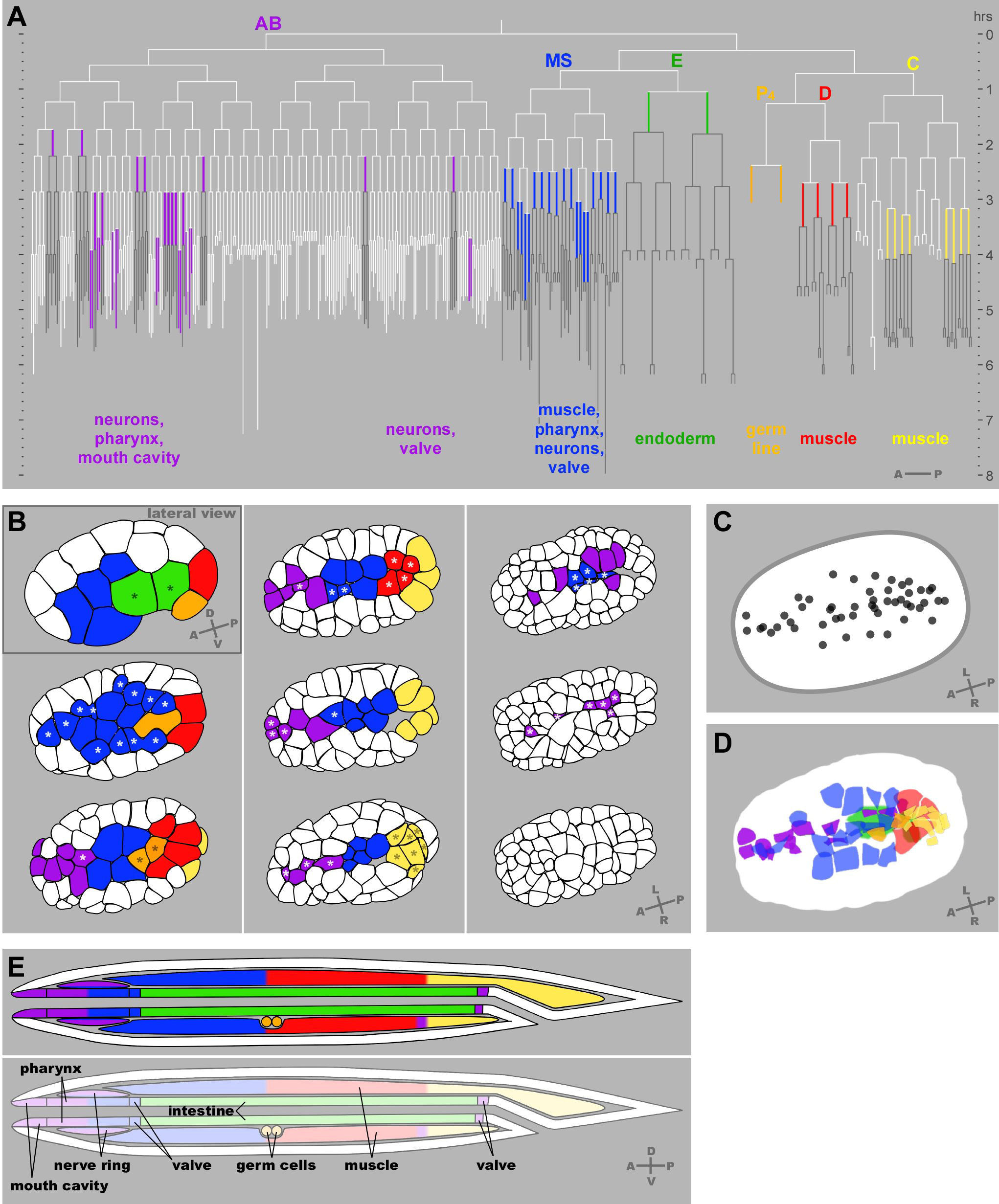 Figure: Gastrulation in C. elegans (click on figure for large format, printable version) (A) Gastrulation mapped onto the cell lineage. The C. elegans embryonic cell lineage with the 66 gastrulating cells each represented in colors here and in (B,D,E) corresponding to the six traditionally-recognized cell lineages. Each vertical line represents a cell, with its length indicating its cell cycle period. Horizontal lines are cell divisions. The anterior daughter at each division is to the left, posterior to the right, as indicated on the lower right. Descendants of gastrulating cells, which generally remain internalized, are drawn in dark gray. Internal structures that each set of cells produces are indicated below. (B) Timepoints throughout gastrulation. The positions of the gastrulating cells are shown in color. Renderings of cell outlines were generated from a spinning disk confocal film of a membrane-marked embryo viewed at a plane through the middle of the top layer of cells, adjusting to match the middle of the top layer of cells as cell divisions resulted in smaller and smaller cells. All views are ventral views except the first stage shown, a lateral view. Here and in the accompanying Movies 1 and 2 in the supplementary material, E, MS, P4, D and all of their descendants are colored from the time they are born. In the AB and C lineages, only some descendants gastrulate. For these lineages, we have colored the AB cells (in purple) only during the cell cycle at which each cell internalization occurs, and we have colored the C lineage (in yellow) at the birth of C, with yellow later marking only those C lineage cells that internalize. Nine embryonic stages are shown, with asterisks marking cells that are about to internalize (that internalize before the next stage shown). (C), (D) The positions from which cells internalize map to a stripe along and near the ventral midline. (C) The center of each cell’s apical domain is marked by a dot three minutes before its internalization was complete, from a ventral view, showing that most cells internalize from a position near the embryo’s ventral midline. 50 of the 66 cells (all but 16 of the AB lineage cells) could be seen internalizing from this view. (D) As in (C), except the entire apical domain is drawn. Endoderm was drawn here based on a separate movie. One cell of the 50 indicated in (C) is not represented in (D). This cell internalized by division toward the interior in this specific embryo. (E) Final positions of the progeny of internalized cells, in the interior of the worm. The antero-posterior order of lineages during gastrulation is mostly maintained through to development of the worm (note the order from anterior to posterior of mostly purple-blue-green-red-yellow in both the gastrulating embryos and the worm). Diagram of a hatched, larval worm (L1 stage, after Sulston et al., 1983) indicates contributions to internal tissues from each lineage. All 66 of the cells above that we observed gastrulating:
|