|

Mitosis and Cytokinesis
in a PtK1 Cell
Stero movie of a mitotic spindle
in a mammalian cell
by Julie C. Canman and Elise Shumsky
Get
out your Red-Green 3-D glasses for this one!
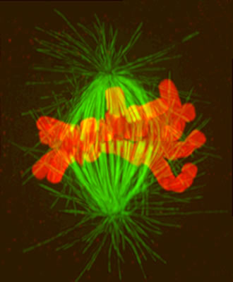
3D
image of a prometaphase cell labeled for DNA and tubulin
by Paul Maddox and Julie C.
Canman
Phase contrast microscopy of M-phase in femxle
rat kangaroo kidney epithelial cells
(PtK1 Cells)Notice
both kinetochores on one chromosome were attached to the same
pole at anaphase onset
by Julie C. Canman and Paul
Maddox
Mitosis and Cytokinesis in a Newt Lung Cell
DIC
microscopy of cell division in a newt lung cell
by Vicki Skeen and E.D. Salmon
Speckled Spindles
Waterman-Storer, C. M., A. Desai, J. C. Bulinski and E. D. Salmon.
1998. Fluorescent speckle microscopy: Visualizing the movement,
assembly and turnover of macromolecular assemblies in living cells.
Current Biology. 8:1227-1230.
Waterman-Storer, C. M. and E. D. Salmon. 1998. How microtubules
get fluorescent speckles. Biophysical Journal. 75:2059-2069
Fluorescent speckle microscopy of metaphase
MT dynamics
or try 2x Zoomed
by Clare M. Waterman-Storer
Fluorescent speckle microscopy of in vitro
mitotic Spindle MT dynamics
or try 2x Zoomed
by Arshad Desai and Clare M.
Waterman-Storer
Anaphase
in vitro
Desai, A. P. S. Maddox, T. J. Mitchison and E. D. Salmon. 1998.
Anaphase A chromosome movement and poleward spindle microtubule
flux occur at the same rates in Xenopus extract spindles.
Journal of Cell Biology. 141:703-713.
Anaphase
in
vitro
by Arshad Desai and Paul Maddox
|