March 24th lecture
Embryology Biology 441 Spring 2008 Albert Harris
Begin with review about nerve pathways and connections
1) Nerve "outgrowth" is really an example of what? ? Secretion of axons? Assembly of short pieces? Or What?
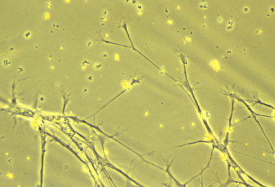
Nerve axon "outgrowth". Locomotion of nerve growth cones, creating axons. These nerves are cultured on a very thin sheet of silicone rubber, for the purpose of detecting the traction forces that cause cell locomotion. But nerve traction is too weak to be detected by this method.
 Mixture of nerve axons and glial cells cultured on a thin layer of silicone rubber
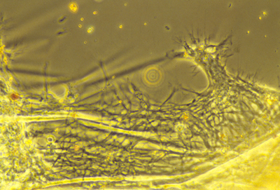
 A motor nerve (vertical) innervating a small muscle inside a living Xenopus tadpole
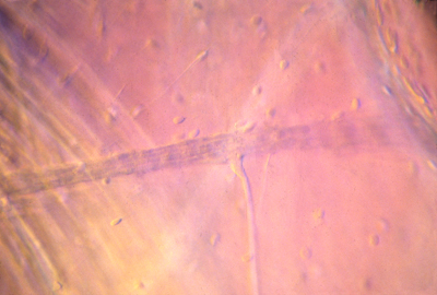
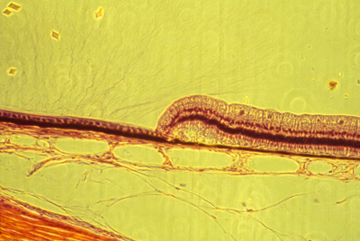
 Bundles of sensory and motor nerve axons often closely parallel arteries, as you will see in anatomy textbooks.
There are two bundles of axons in this histological section, one in the lower left corner and the second close above it, among fat cells.
Can you find the artery wall on the right?
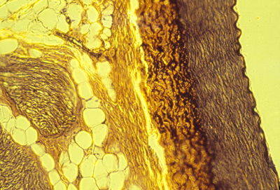
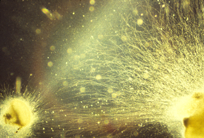 Nerve "outgrowth" can be guided (somewhat!) by aligned fibers of collagen. In this photograph, hundreds of nerve axons are "growing" (crawling) toward the left and upward from a piece of nerve tissue at the lower right corner, through a gel of re-precipitated collagen.
David Stopak and I had hoped to discover much more alignment of nerve axons along the line of oriented collagen fibers that extend from the mass of mesenchymal cells at the lower left toward another mass of mesenchymal cells just outside the field of view off the top center of the photograph. A famous German scientist named Paul Weiss had hypotehsized that oriented collagen fibers should be a "Nervenbahn" (like an Autobahn for nerve growth cones). Unfortunately, it doesn't seem to work very well.
2) How was the true cause of nerve fiber extension proved? Approximately when? But it wasn't done by a UNC faculty member!
3) What very important method was invented for
the specific purpose of proving how nerve fibers extend?
(and approximately what year was this discovery/invention made)
[1907]
4) Did you know that this method was the key breakthrough
in the development of vaccines against polio & some other diseases?
Figure out why. Discuss. Ask.
5) Do nerve axons actually get attracted toward certain places?
(What force does the "pulling", if any.)
6) When our textbook talks about nerve axons being "pushed" away from certain places during embryonic development, does Scott Gilbert literally mean there is a pushing force, or what?
Maybe a weakening of the pulling ability of nerve growth cones?
7) What is meant by a "neural projection"
8) Are there "projections" of motor nerves,
as well as projections of sensory nerves?
Sure.
9) What about projections of nerve connections
Between one part of the brain and another?
(yes, lots of them)
10) Which neural projection has been the subject of the most experimental research?
The Retino-Tectal Projection!
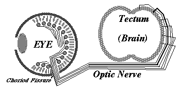 Diagram of the pattern of innervation of the optic tectum by axons from the ganglion cells in the innermost layer of the neural retina of the eye on the opposite side of the head
 section of neural retina
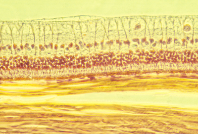
 Lateral edge of the neural retina in a histological section through a rabbit eye. In the left half of this photograph, the neural retina is just a thin epithelium one cell layer thick, and only about twice as thick as the (black) pigmented retina just below it. About the center of the photo, rather abruptly, the neurfal retina has developed all its layers of nerve cells.
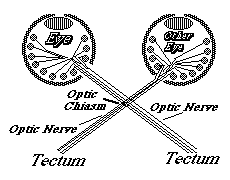 Diagram of the innervation of the optic tectum of the brain by nerve axons that crawl from the ganglion cells of the eyeball on the opposite side of the head.
 Diagram of a neural projection. Nerve fibers from one area (such as the retina) crawl so as first to form a bundle, and then spread out to connect to some specific part of the brain in the same (map-like_ geometric pattern as the places from which the nerve fibers started. For some unknown reason, the retina-brain connections pattern is upside down and backwards (and so are som other neural projections).
&&&&&&&&&&&&&&&&&&&&&&&&&&&&&&&&&
Some Categories of mechanisms by which nerve axons can be guided:
a) "Contact guidance" nerves and other crawling cells will line up along fibers, grooves and ridges.
b) Adhesion gradients "Haptotaxis"
c) "Chemotaxis" and also "negative chemotaxis"
An important medical problem is how to guide nerve axons Back to their normal points of connections, to form synapses.
Many variations of this problem arise in surgery, neurorology, etc.
IMPORTANT NOTE: (expressed as a thought question)
Which of the following will cause chemotaxis:
Maybe ALL of them
I) If the right and left front edges of each nerve growth cone detect the concentration of some particular chemical, and then turns toward the direction where the higher concentration is detected?
II) If the front of the nerve growth cone keeps measuring the concentration of a chemical, and keeps going in the same direction as long as this chemical concentration continues to increase with time, but makes a random turn whenever the concentration of the substance begins decreasing?
III) If the front of each nerve growth cone detects a chemical concentration, and some part of the nerve fiber behind the leading edge also detects the concentration of this same chemical,..
and the growth cone keeps crawling in the same direction as long as the chemical is more concentrated at the front that behind the front�
But retraction, and a random turn, occurs whenever the concentration behind the front is higher than the concentration of the chemical at the front.
IV) If the adhesiveness of a nerve growth cone increases in proportion to the local concentration of some diffusing chemical.
V) If forward crawling of nerve growth cones Becomes slower, or stops completely, whenever The local concentration of some diffusible chemical Is detected to be larger that a certain threshold concentration?
Which of these will seem like positive chemotaxis? (Or do all five of these kinds of mechanism deserve to be called "chemotaxis"?
Or maybe: "Type One Chemotaxis" "Type Two Chemotaxis" etc.
Experimentally, for example when studying nerve cell paths in an embryo, are you going to be able to distinguish which form of chemotaxis is causing guidance, (or if any of them are)?
Maybe you can invent some other possibilities?
Could nerve guidance be by some completely different kind of mechanism?
************************************************
When you study embryological pattern formation (such as the connections of nerves to form a brain) can you distinguish which alternative mechanism causes each part of a pattern?
(first you need to invent as many as possible different alternative kinds of mechanism?
Or should you just say "pull" and "push" and "grow", and "chemotaxis" (as if there were only one kind!) and hope for the best.
Unfortunately, that's been what most researchers (and nearly all textbook writers) have done.
************************************************
A more specific question:
What control mechanism causes formation Of the retino-tectal (optic nerve) projection to the brain?
Steve Roth proposed a theory of adhesion gradients. He discovered that cells from the upper half of the retina were more adhesive to the lower half of the optic tectum.
He proved this using chicken embryos by dissociating halves of retinas into individual cells. (very much like Wilson, Holtfreter, Trinkaus had done)
He dissected out early chicken mid-brains, kept them in tissue culture medium, stuck by pins to the bottom of culture dishes,
And let single cell suspensions of retinal cells settle onto these pieces of brain, then gave them an hour to form adhesions, washed off the retinal cells that hadn't stuck, and counted how many retinal cells had stuck to different parts of the midbrain.
The result was that about twice or three times as many cells of the top half of the retina stuck to the lower half of the optic tectum, and vice versa.
In other words, more retinal cells stick (more strongly?) to the half of the roof of the mid-brain that would have been innervated by ganglion cells from that part of the retina.
This, it seemed that the mechanism of neural projection formation seemed to be some kind of gradient of cell-cell adhesion proteins or other molecules.
^^^^^^^^^^^^^^^^^^^^^^^^^^^^^^^^^^^^^^^^^^^^^^^^^^^^^
Since then, evidence has been discovered that what happens is more like negative chemotaxis.
The ganglion cell axon growth cones are attracted toward the correct general area of the brain, but then their adhesion is inhibited by a gradient of a protein called ephrin. Ganglion cell surfaces have specific receptors for ephrin.
And there is a gradient of amount of ephrin receptors on ganglion cell surfaces, combined with a gradient of amounts of ephrin on brain cells.
Plus there are two kinds of ephrin: ephrin A and ephrin B.
each with their own kind of receptor.
The ephrin A gradient is perpendicular to the ephrin B gradient; likewise there are 2 perpendicular gradients of ephrin receptors.
The key idea is that the more ephrin receptors a cell has, the more strongly its axons will be "repelled" by any given amount of ephrin.
Likewise, the parts of the brain surface with the most ephrin will more strongly repel the axons (per amount of ephrin receptor).
Therefore: ganglion cell axons with the smallest amount of receptors will be least inhibited or repelled, and therefore will extend farthest up the gradient of ephrin concentration.
Ganglion cells with the most ephrin receptors will be the most sensitive to ephrin, and will be stopped at the low end of the ephrin gradient.
Note the unstated assumption that some other "attraction" mechanism is guiding the ganglion cell nerve growth cones "upward" along the gradient of (inhibitory) ephrin.
Another protein that specifically serves to stimulate retraction of crawling nerve cell growth cones has been named semaphorin.