January 23, 2008 lecture
Embryology Biology 441 Spring 2008 Albert Harris
Active Cell Movements That Form the Embryonic Body
1* Invagination of Epithelial Cell Sheets (= active folding of epithelia) driven by actomyosin contraction localized at whichever surface becomes concave during the folding; although there is evidence for some other mechanisms)
examples:
Gastrulation in sea urchins
Neurulation in mammals, birds and amphibians
"outfolding" of kidney, lung, salivary glands etc.
-
Neurulation in chick sections
2* "Amoeboid" Cell Locomotion.
Individual cells, and also the edges of cell sheets, crawl by cytoplasmic acto-myosin exerting "traction""= as a shearing force on collagen, glass, rubber, etc.
The directions, strengths and locations where cells exert traction are studied by putting tissue culture cells on thin sheets of rubber or on gels of fibrin, collagen, or artificial chemicals (acrylamide)
-
A single cell on a thin sheet of rubber
Cell traction can also pull collagen past cells, either to compress collagen or to align it at distances as far as centimeters
-
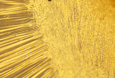
Collagen around cartilage
Most textbooks say that tissue cells exert traction and crawl
by reaching out flat lamellipodia or thin filopodia in front
of the cell, then sticking these protrusions to the collagen (etc.)
and then contracting the protrusion, so as to pull the cell forward.
THIS IS NOT REALLY HOW BODY CELLS MOVE.
I have studied it for years, and never seen any such "reach-grab-pull".
But lots of otherwise good scientists believe it, so in future courses and standardized tests, this fiction may be the "right" answer!
++++++++++++++++++++++++++++++++++++++++++++++++++++++++++++++++++++
Epithelial cells form continuous sheets, without gaps.
(They can hold back flow of liquids)
Some epithelia are one cell thick, such as those that line blood vessels.
Other epithelia are many cells thick, such as the epidermis which is the outer layer of our skin.
Mesenchymal cells crawl around as individuals, and sometimes in groups, and can also form sheets, but with gaps.
Also, many epithelial cells can crawl as individuals But this wouldn't occur normally, except in wound healing. But epidermal cells of fish and amphibians crawl very fast as individual cells, which lots of researchers study, and call "keratinocytes".
These crawl at a very steady (comparatively fast) speed, almost in straight lines, although the elongate perpedicular to their direction of movement, and exert "sideways traction" forces, as was discovered by Prof. Kenneth Jacobson in the UNC Med School.
These cells are easy to get: just use forceps to pull individual scales from a fish,
gently disinfect the scales, separate the cells with enzymes, or just
late the cells out in tissue culture.
(This need not harm the fish, which can be put back into pond or aquarium;
Fish frequently bite out scales from each other in fights)
I suspect this unusual pattern of cell locomotion is "meant" to constrict and close penetration wounds. Such wounds close within minutes in fish and frogs, by means of a combination of contraction along the edge of the wound, plus crawling traction toward the center of the wound.
When single isolated cells exert this same combination of sideways contraction And forward traction, the result is fast & steady locomotion.
The British-Austrian researcher Victor Small discovered a flow of cytoplasmic actin rearward near the bottom of these "keratinocytes" and forward near the top of their cytoplasm. Two friends and I wondered if this could be true, and I provided some fish skin cells, which they injected with fluorescently-labeled actin, and observed on a very special microscope; and it was clear that Prof Small was absolutely correct about the pattern or actin flow, like a tractor tread.
Critics often doubt whether scientific discoveries really get repeated and checked. I believe they do; and this is one of many examples. If our results had contradicted the published ones, then we would have written to the author, and suggested what might have gone wrong. Eventually, he would have published a correction, or we would have published a correction.
-----------------------------------------------------------------------------
Other kinds of morphogenetic cell movements:
* Conversion of epithelial cells to mesenchymal cells.(This doesn't have a special name, like "invagination" but it deserves one: many important tissues form this way)
Examples:
1) Formation of the "Neural Crest" ectoderm (by the cells along the edges of the neural plate, where its edges fuse together.)
Neural crest cells later differentiate as the following:
(Which you should memorize)
-
a) Sensory nerve cells (that connect to all your skin, except on the head)
b) Autonomic nerve cells (the postganglionic ones)
c) The medulla of the adrenal gland. (really = autonomic nerves)
d) Pigment cells of the skin (but not those of the retina)
e) Schwann cells (that form myelin sheaths around nerve axons)
f) Skeleton of the facial area! (but the rest of the skeleton is mesoderm)
g) "Odontoblasts": The special differentiated cell type that forms
the inner part of the teeth.
Note that research on neural crest is
heavily concentrated among the faculty of Dental Schools, especially UNC's.
h) A few other special cell types, some not discovered yet.
Embryologists have long wondered and argued why so diverse A subset of cell types should all develop from neural crest.
That's a good idea, but obviously leaves much unexplained.
The neural tube is the only example I know of where mesenchymal cells are released where epitheila sheets fuse, although there are many, many other examples of epithelial fusion.
How did researchers discover +/or prove that these diverse cell types
are all formed by differentiation of neural crest cells?
Mostly
by grafting cells from embryos of Japanese Quail into (early) embryos
of chickens, to make quail-chicken chimeras.
It so happens that the Japanese species of quail have nuclei that stain
very differently from chicken nuclei; so that in histological sections,
you can tell them apart easily and without doubt
By carefully grafting just the neural crest cells, scientists mostly in France,
proved exactly which organs and cell types come from neural crest;
and the same method also works on any other part of embryos.
Earlier research had used radioactive labeling of DNA, followed by Grafting neural crest from one embryo to another. (1950s)
Before that, and since, scientitists grafted neural crest from eggs of one strain of chicken to another, and showed that their feather color patterns change according to the source of neural crest
In a 1940s experiment by my PhD thesis advisor, they actually
got a Robin's egg out of a nest, and grafted some of its neural
crest into a developing chicken egg. Guess what happened!
Yes, when they hatched the eggs and raised the chicks,
the adult chickens developed grey backs and orange fronts.
Other important examples of epithlial cells becoming mesenchymal cells:
2) The " Primary Mesenchyme" of sea urchin embryos, formed early in gastrulation.
These cells later secrete the skeleton of the sea urchin larva = " Pluteus"
3) The " Secondary Mesenchyme" of sea urchin embryos, formed later in gastrulation;
These cells later differentiate into muscle cells of the Pluteus larva
4) Gastrulation in mammals, birds and reptiles occurs by release of epithelial cells
from what had been continuous epithelial cell sheets.
The cells that make up these particular epithelial cells sheets
are called " epiblast cells"
Below them, between then and the yolk,
is a thinner sheet of epithelial cells called " hypoblast cells"
A thickening of the epiblast cells occurs along what will be the
anterior-posterior axis of the bird, reptile or mammal body.
This thickening is called the " Primitive Streak".
Down the middle of the primitive streak, many of the epiblast cells change into mesenchymal cells, and after they have made this change they are called " mesoblast cells"
Most mesoblast cells later differentiate into mesodermal organs, But some mesoblast cells differentiate into endodermal organs!
Hypoblast cells differentiate into endodermal tissues that are part of extraembryonic membranes (placenta in mammals). Supposedly, none of the hypoblast cells make it into any Part of our digestive tract, lungs, liver etc. endodermal organs of the body.
Another important category of morphogenetic cell movements:
* Conversion of mesenchymal cells into epithelial cells.
Usually, this results in forming some kind of enclosed cavity, lined by the new epithelial cells.For example, mammals, birds and reptiles form their coelomic cavity by what had been a broad sheet of mesoblast cells (mesenchymal) converting into two sheets of epithelial cells, facing each other, so as to form a liquid-filled cavity between them (the "Coelom').
The term "cavitation' is often used to refer to examples Of conversion of mesenchymal cells into epithelial sheets. (Why? Can you guess?)
Other examples of conversion of mesoblast cells into epithelia:
* Formation of kidney tubules. * Formation of capillaries, and later arteries and veins * Formation of somites (segmental blocks of tissue, that control the locations where ribs, vertebrae, segmental nerves, and other segmental structures will form, later in development.
Please notice that the somite cells soon re-separate into mesenchymal cells, and that many of the segmental structures whose locations they control develop from ectoderm, or from non-somite mesoderm; Nevertheless, and by mechanisms yet to be discovered, where the somites form controls where the segments form. (Example: vertebrae of your back-bone; you develop one vertebra for each pair of somites.)
Questions for class discussion:
a) What would you expect to happen if 5 small somites from the tail of a salamander embryo were grafted in place of 3 (larger) somites from the trunk area of the same or another salamander embryo.b) How do you think scientists discovered that somites control where ribs etc. form, and how many bones you develop in your backbone?
c) What alternative kinds of mechanism could control where somites form?
Two (?) more important kinds of morphogenetic cell rearrangement.
* Fusion of two epithelial cell sheets, to form one continuous sheet.Remember the neural plate folding its edges together?
Fusion of these edges creates :
-
1) a continuous neural tube
2) a continuous somatic ectoderm
Examples:
a) Spina bifida (incomplete fusion of the edges of the neural tube).
b) Cleft palate (failure of fusion of two structures that form the roof of the mouth).
c) "Hare lip" (Cleft lip) (incomplete fusion of sheets of tissue that form the face).
** Because epithelial sheets have two very different kinds of surfaces:
The basal surface, which faces other tissues, mostly mesenchymal.
The apical surface, that faces fluids or the outside world, and may have flagella.
Many scientists think there must be fundamental differences between epithelial fusions at apical surfaces, and those at basal surfaces.
Examples: of basal-basal fusions
Connection of one capillary to another.
Connection of the mouth to the rest of the digestive tract.
What's an example of an apical-apical fusion?
That was just discussed above?