January 24-29, 2007
Embryology Biology 441 Spring 2007 Albert Harris
Organs that form from Ectoderm
Neural Tube Ectoderm --> brain, spinal cord, motor nerves, retina

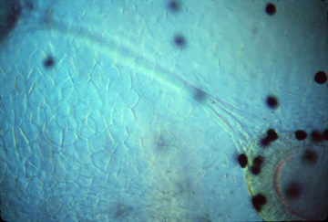
In this section of a chicken embryo, you can actually see the neural crest separating from the neural tube below and the somatic ectoderm above and to each side. (Also notice how small the notochord is, under the neural tube).
Neural Crest Ectoderm --> sensory nerves, pigment cells, Schwann cells, (post-ganglionic) autonomic nerves,
adrenal medulla, skeleton of face, and odontoblasts (dentine)
(and several other kinds of cells, too; including some in the heart)
Somatic Ectoderm -->
-
epidermis (the outer layer of the skin)
hair
feathers
scales in reptiles, birds & mammals
ameloblasts (form enamel of teeth)
{Except shark scales are very much like teeth! I don't know if they are formed by a combination of neural crest & epidermis?}
Somatic Ectoderm--> Stomodeum (infolding that forms the mouth)
Part of the roof of the stomodeum forms "Rathke's Pocket" which differentiates to form the anterior pituitary gland.
At least some of the salivary glands form as out-foldings of the innermost parts of the stomodeum.
Also formed by cells of the stomodeum are ameloblasts
These secrete the outer, enamel, layer of teeth
They are induced to form by the odontoblasts, which develop from neural crest cells.
This is an example of reciprocal induction, since both cell types apparently induce development of the other.
(neither can develop without the other)
Prof. William Koch of the UNC med school did some of the most important early research on this reciprocal induction)
*******************************************************************************************
The (previously famous) Kollar and Fisher Experiment
They grafted mouse odontoblasts to pieces of chicken stomodeal epithelium, and cultured these in the aqueous humor of the eye of mice! Yuk!
They reported the formation of teeth, with enamel secreted by the chicken cells. Although no bird has had teeth for 50 million years!
The early birds, that lived in the age of dinosaurs, had teeth.
But beaks are lighter, which is probably why selection favored mutant birds that didn't form teeth.
Which genes could have been inactivated or deleted?
-
a) Genes for enamel?
b) Genes for differentiation of odontoblasts?
c) Genes for receiving inductive signals?
d) Genes for sending inductive signals?
etc.) Can you invent some other possibilities?
Which alternatives do those results seem to disprove?
Why would you guess that these experiments have been dropped from recent editions of embrylogy textbooks? Can you invent some possible mistakes or serious misinterpretations that could have been made?
What if some mouse ameloblasts were accidentally included in the grafts? Or what if some mouse odontoblasts had redifferentiated into ameloblasts? What further information could prove that K & F's interpretation was correct? Or not?
A little more about the epidermis:
Germinative layer:
a layer of actively dividing, undifferentiated (but determined) cells (a good example of what "stem cells" used to mean)
cornification: filling of the cytoplasm with a protein called keratin. This makes them impermeable, so that they die.
(note that this cell death is completely different from apoptosis)
Hair, finger-nails, claws, scales and feathers are all made of highly cornified epidermal cells.
hair: formed entirely by somatic ectodermal cells (epidermis)

But each hair's formation is induced by a tightly bunched-together mass of mesodermal mesenchymal cells, called a dermal papilla. These control where hair form, and (apparently) the diameters of each hair.
Feathers in birds are also induced to form by mesodermal papillae. Also scales in reptiles, birds and mammals, also each have a dermal (mesoderm) placode.
The locations of feathers and scales is EXTREMELY regular, almost crystalline in pattern. It is a long standing unsolved problem to discover the mechanisms that controls where they form

Hair locations are usually much less regular, except for certain hairs like the vibrissae above the upper lips of dogs.
Also, the next time you are petting a dog, notice that the formation of regular-sized hairs seems to be inhibited for a short distance around each of the vibrissae.