with a portrait of H.V. Wilson, UNC Department of Biology. Professor Hairston was H.V. Wilson's last graduate student.
| Oil painting of Albert Harris by his father Kenneth Harris, painted about the time the future Professor was first getting interested in Biology. | 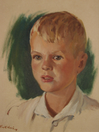 |
|||||||||||||||||||
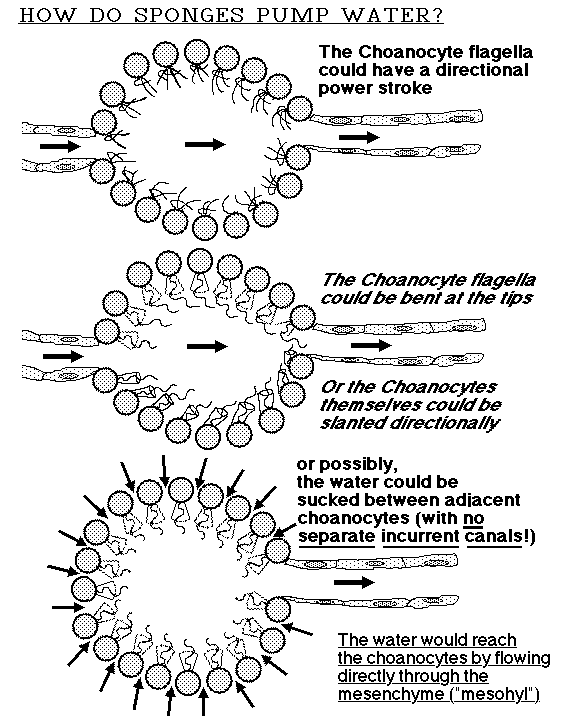
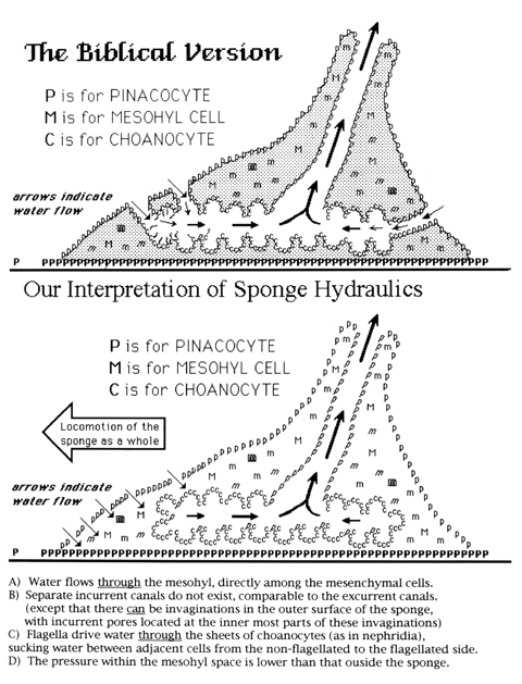
| Bond and Harris also discovered that small sponges crawl (instead of being sessile, as textbooks still claim), as well as that the geometrical pattern of flow and propulsion of water through the interior of sponges is very different from what the textbooks say. This flow is analogous to the flagellar pumping of water through the category of invertebrate kidneys called nephridia; in other words, water is sucked from the mesohyl space between flagellated choanocytes, instead of being pushed along past the apical ends of choanocytes, as has always been assumed. Excurrent canals really do exist, but are closed at the end from which the water is being pumped. No incurrent canals lead to these excurrent canals, however, except to the extent that indentations sometimes occur in the outer surfaces of sponges. Water is pulled inward through holes in a special kind of surface cell, and then flows through the same "mesohyl" spaces where the spicules are located, before being sucked between choanocytes into the excurrent canals. |
 |
 |  |
|
 |
 |
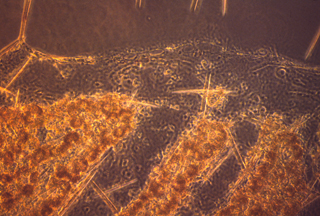
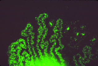
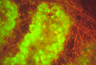
| This pattern of water flow can be made visible by feeding small particles to sponges, or by feeding them fluorescent beads, or by dissolving fluorescent dyes in their water. |