L.P.M. I.M. Somites Notochord Somites I.M. L.P.M
We are members of the phylum "chordata", all of the members of which have a notochord at some stage of their development.In mammals and birds, the function of the notochord is replaced by the vertebral column (bones of the back-bone+intervertebral discs)
But tadpoles and larval fish actually depend on the notochord to allow them to swim by alternating contraction of muscles along each side.
The notochord mesoderm also induces formation of the neural tube.
(and it probably is also needed to elongate mammal embryos)
Anyway, our embryos can't develop without a notochord.
The structure of the notochord is a long stack of (not always) flattened cells, that often have very much the geometry of a stack of coins.
Wrapped tightly around the surface of this long cylinder of cells are layers of fibers of type I collagen in many spiraling layers, that alternate in direction (a clockwise layer, then a counterclockwise layer, then another clockwise layer, etc. etc. etc.)
The cells inside the notochord then form large vacuoles in their cytoplasm, which makes each cell swell in volume, pushing against the spiral layers of (strong & non-stretchable!) collagen fibers. The result is that the notochord can bend side to side,
but can't be compressed along its anterior-posterior axis.
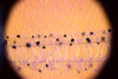
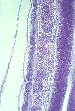
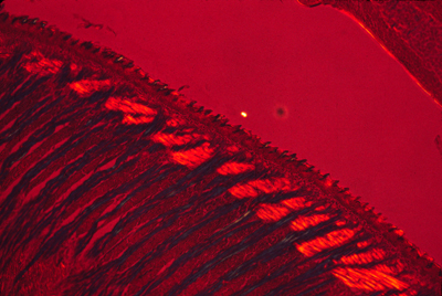
Tail of a living tadpole. The broad line just below the center
is the notochord, and you can see that it is filled with highly vacuolated
cells. The neural tube is above it. You can also see pigment cells and
mesenchymal cells scattered around.
A short digression about the VERY important protein, collagen.
Type I collagen is the main extracellular protein of the body, and causes the mechanical strength of tendons, skin, organ capsules, the wall of your eyeball (including even the cornea!).(even bones are 1/3 type I collagen).
Jello (flavored gelatin) is type I collagen; so is Elmer's glue.
Leather is mostly type I collagen that has been chemically cross-linked (this chemical treatment process is called tanning).
Collagen can be purified from rat tail tendons, and skin of cows,
and plastic surgeons sometimes inject it to make skin smooth.
A series of very similar proteins are named "type II collagen"
type III collagen, type IV collagen, and guess what comes next?
With numbers on up into the 20s, each with special properties.
There is more type I collagen in the body that all the rest put together.
For many years, it was an unsolved problem how type I collagen gets arranged into so many different geometric patterns in the body .
Everyone expected that different proteins would be used to make different geometric patterns.
In the early 1980s, a UNC grad student and I did experiments that (maybe?) gave the answer to this paradox. We said that mesenchymal cells pull strongly on collagen, and mechanically rearrange it according to what forces are exerted at each location.
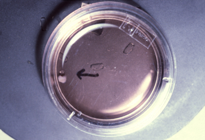
Photograph of a rat collagen gel compressed by chicken cells
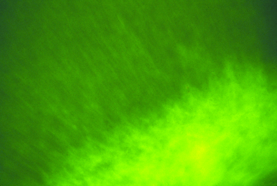
Photograph of fluorescently-labeled rat collagen
When fluorescent collagen was injected into developing chicken embryos, then it got made into many different anatomical structures:
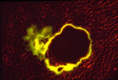
Photograph of chicken artery, with fluorescent rat collagen wall.
Next, back to questions about how the notochord forms.
The best research about this has been done by Ray Keller,
(Currently the chairman of the Biology dept at UVA)
(Also two women bio-engineers, Mimi Koehl and Dany Adams)
They describe a process of convergent extension, in which the future notochord cells extend processes sideways; these then adhere to each other, and these processes actively contract, pulling in the sides.
They think the collagen sheath gets made later, and does not participate in the actual formation of the notochord.
(and probably they are right about that; but I would bet they aren't)
Several odd facts about the notochord:
In reptiles, birds and mammals, the notochord cells are the last to leave the surface (epiblast) at the Hensen's Node.
* In some species (humans & turtles), Hensen's node is partly an invagination, & a hollow tube runs down the middle of the notochord
(this tube then disappears, as the cells push together)
* In sections of amphibian embryos, you can see pigment granules tend to accumulate down the middle of the notochord.
* The spiral layers of collagen fibers that are the sheath of the notochord are oriented at 90 degrees to each other.
This is one of several examples in which collagen fibers mysteriously become oriented in alternating layers, with the fibers of each layer exactly perpendicular to the fibers on the layers above and below.
Also, there are many examples in which muscle cells become lined up in perpendicular alternating layers: Mammal tongues, elephant trunks,
squid tentacles, and in Hydra, both the body and the tentacles.
Prof Bill Kier of this department specializes in studying them.
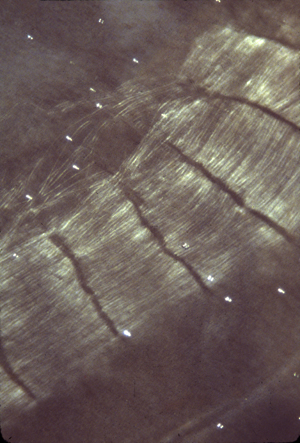
Polarization micrograph of a section through a mouse tongue
Not only are there about 26 muscle layers oriented in alternating directions, but the bright red areas are sections of a third set of about twelve muscles that are perpendicular to both the other layers
Anybody who could figure out the mechanism that creates these perpendicular layer patterns, and prove their theory true, would go down in biological history.
Somites are segmental blocks that form beside the neural tube.
At first, there are continuous columns of "paraxial" mesoderm
Then these columns spontaneously split apart, one pair at a time.
As if a quonset hut were to split into a row of igloos.
Or as if a long piece of french bread were to slice itself.
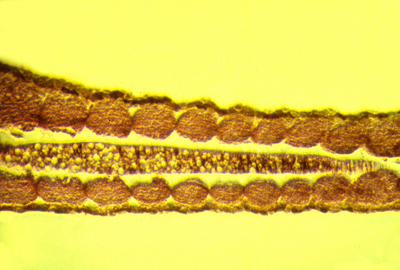
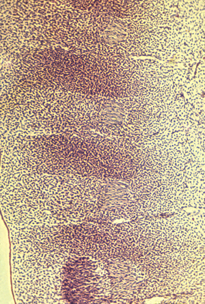
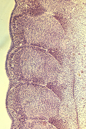
Longitudinal section of notochord and somites in a developing frog embryo
The physical mechanism of splitting apart could be active constriction, or could be decreases in cell-cell adhesion proteins.
(or could be the same as whatever forms feather papillae!)
In scanning EM photos, "somitomeres" can be seen, where a somite is going to form, apparently as the first stages of somite formation.
In most species, somites start as hollow epithelial balls,
and conversion of cells from being mesenchymal to epithelial
therefore seems to be part or the process of separation.
(These epithelial cells seem to have basal surfaces outward)
Much research has been done on the genes needed for somite formation
see pages 467 to 470 in the textbook.
Suspected genes include "Notch", "Lunatic Fringe", and "Hairy1"
The most popular categories of theory are
1) That somite segmentation has the same mechanism as formation of segments in flies, using genes analogous to "even-skipped" etc.
2) The "Clock-and-Wavefront Hypothesis", according to which one quantity oscillates higher and lower in amount, while another variable forms a gradient that gradually increases in amount along its length.
A somite is supposed to be split off each time the oscillator increases.
Really, no one even knows whether we should think of the splits as being the real entities, as opposed to the somites themselves.
For most species, the same number of somites forms in each embryo.
(the number is 32-34 in humans, 50 in chicks, 65 in mice (many in tail)
more than 500 in some snakes, and varies with temperature in fish!
You form as many vertebrae as your embryo had formed somites.
Each somite subdivides into four parts: