Building a Blood Vessel is Hard Work

Welcome to the Bautch Lab
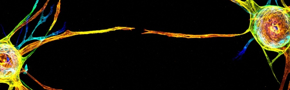
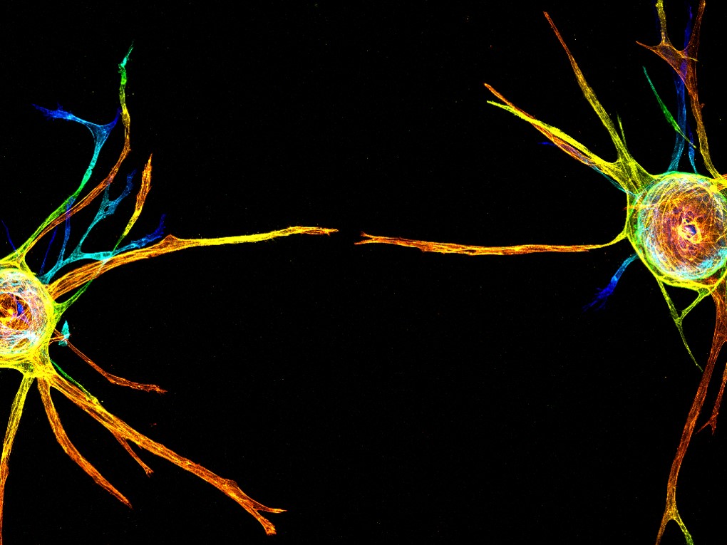
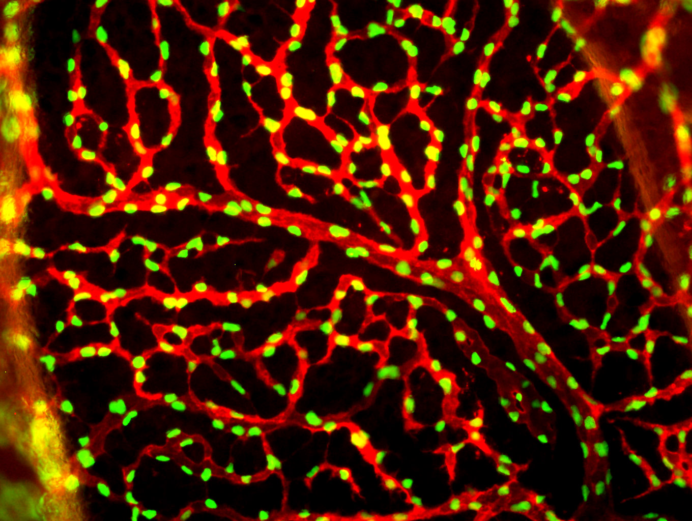
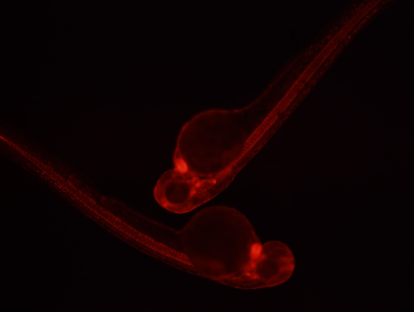
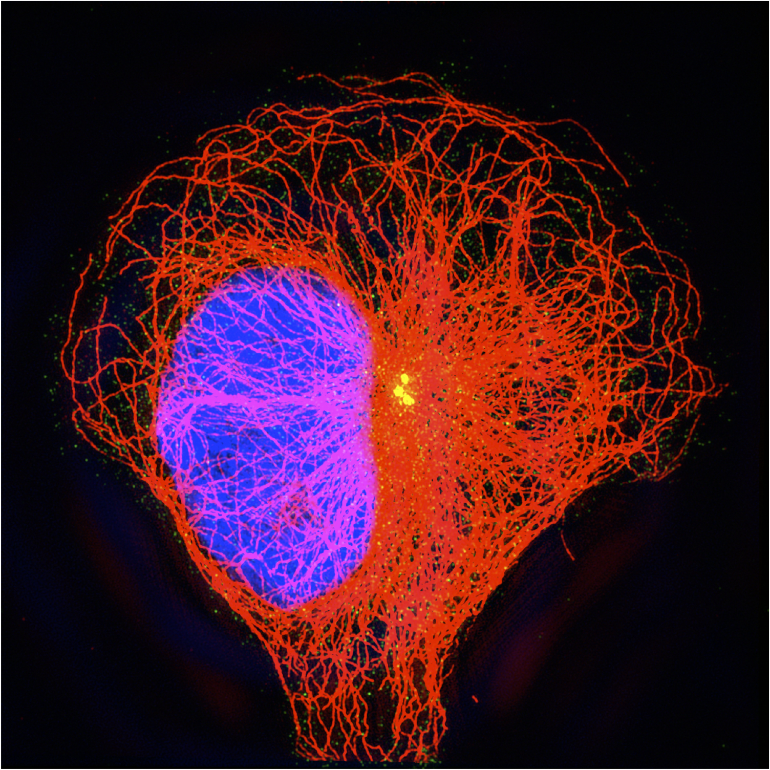
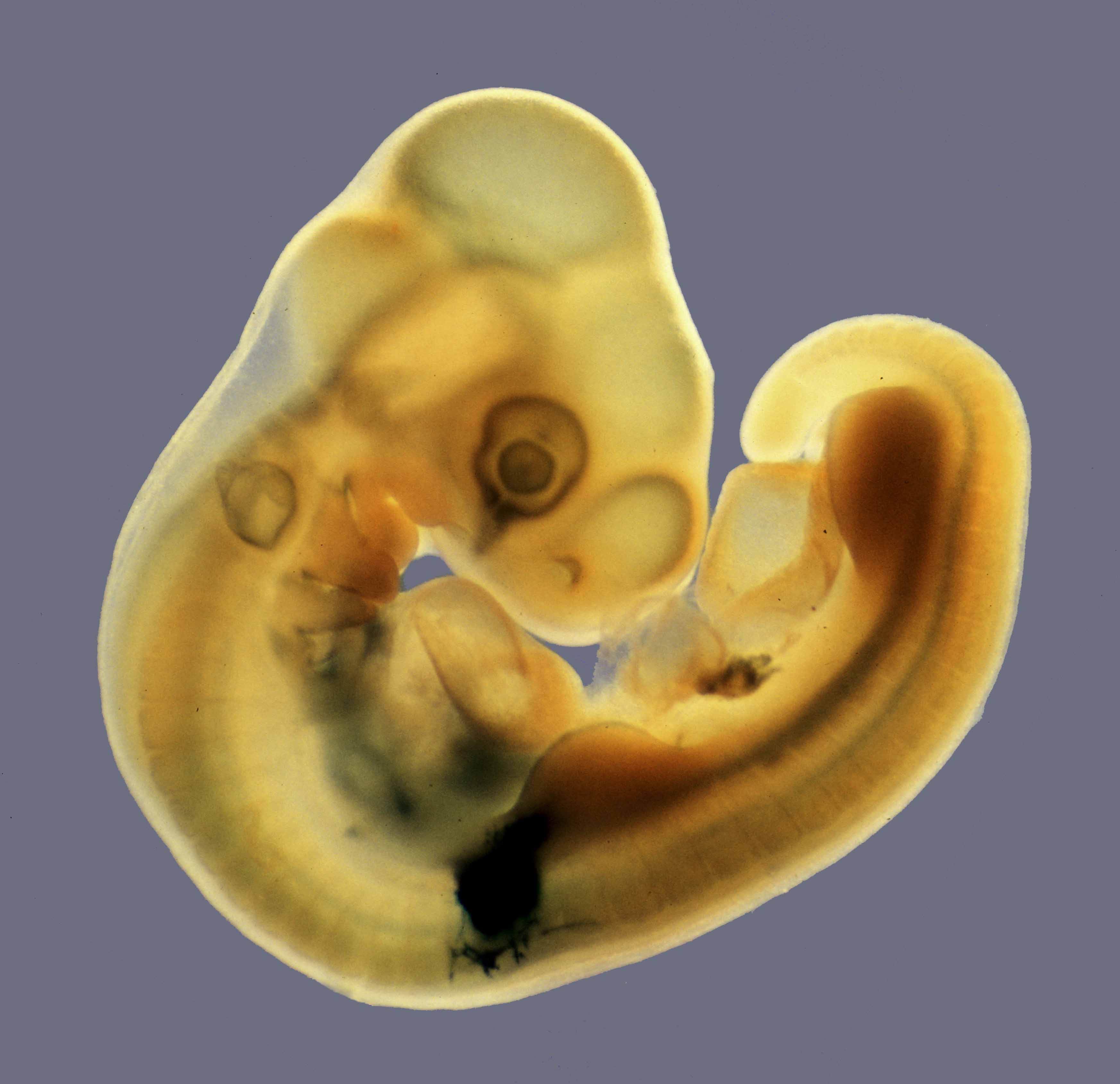
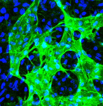
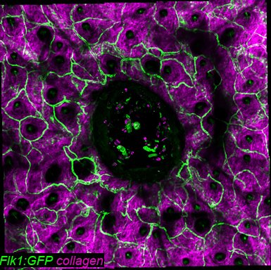
Research in the Bautch Lab centers on the molecules and processes that control development and disease. Our major focus is the study of how blood vessels form and are patterned during development, and how these processes are disturbed or co-opted during diseases such as cancer. The group of scientists working in this lab include graduate students, post-docs, research technicians, and undergraduates.